Casey DEAN, Kelli SORBY
In 2019, Casey Dean presented at the Fertility Society of Australia and New Zealand’s Scientists in Reproductive Technologies conference. Their talk was on Filaments, slender, thread-like structures that play crucial roles during embryonic development. These filaments can be found in various cellular components and processes involved in embryonic growth and differentiation. Here are two important examples of filaments in embryology:
- Actin Filaments: Actin filaments, also known as microfilaments, are thin, fibrous structures composed of actin proteins. They form a part of the cytoskeleton within cells and are involved in numerous cellular processes, including cell division, cell migration, and cell shape changes. During embryogenesis, actin filaments provide structural support to the developing embryo and contribute to important morphogenetic events such as cell differentiation, gastrulation, and neurulation.
- Microtubules: Microtubules are long, hollow filaments composed of protein subunits called tubulins. They also form a part of the cytoskeleton and are involved in a variety of cellular functions. In embryology, microtubules are essential for processes like mitosis (cell division), intracellular transport of vesicles and organelles, and establishment of cell polarity. Microtubules also have a role in determining cell fate and forming the scaffold for
the development of the nervous system.
During their research they had observed string-like structures appear during first division looking like they were “pulling fragments” out of the embryos. They named these structures as filaments due to their appearance, as yet they hadn’t found any reports about them.
They asked:
- Does the amount of filaments correlate with the degree of fragmentation formed at first division?
- Is filament formation related to the location of the cleavage furrow relative to the cell-zona anchoring?
- Did filaments, and subsequent fragmentation formation, affect clinical pregnancy rate?
During this research, 1555 embryos were assessed.
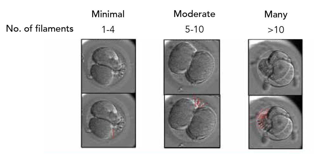
They found that filaments were observed in 70% of the embryos. Of those 70%, they found an even spread of filament volume, between minimal, moderate and many, the occurrence of filaments was even. There seems to be no correlation with the volume of filaments and the degree of fragmentation.
Unsurprisingly most embryos that exhibited filaments were significantly more likely to produce clustered fragments. In embryos with comparable degrees of fragmentation, clinical pregnancy rate was significantly lower in embryos that exhibited no filaments.
What our scientists believed may be causing this is that their fragmentation is caused by abnormal fragmentation, like that blebbing from earlier, which is more likely to indicate apoptotic processes rather than a mechanical feature.