Pelvic Ultrasound
Screening Tests at No.1 Fertility
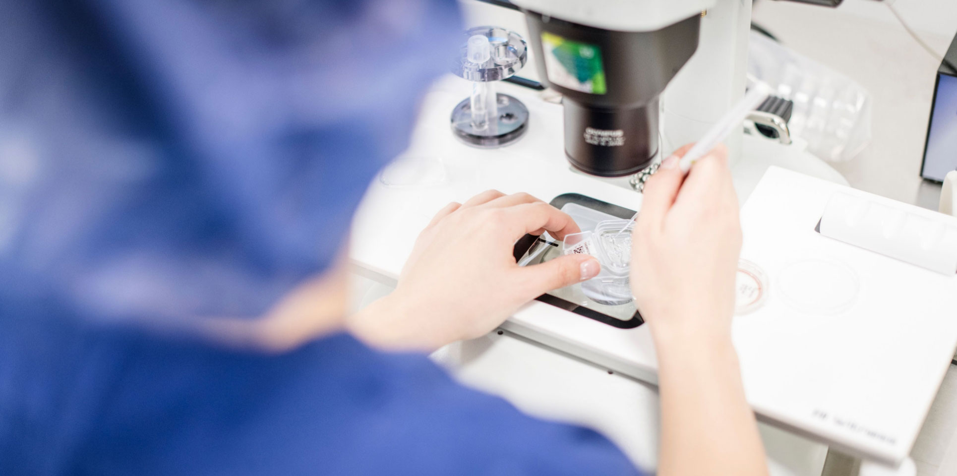
At No.1 Fertility, we believe preparation is key to success. Before starting your treatment, our comprehensive screening tests help give your specialist a clear picture of your reproductive health. These tests are an essential first step to ensure the safest, most effective treatment journey possible.
Our specialists use your screening results to tailor care specifically to you — because at No.1 Fertility, every detail matters when it comes to achieving your dream of parenthood.
Screening may include blood tests, ultrasound scans and semen analysis, depending on your treatment plan. Please note that your doctor may request additional tests as needed. Screening blood tests must be completed before the start of your cycle.
Screening for Egg Providers and Gestational Carriers
If you’re providing eggs or acting as a gestational carrier, your screening helps us check both your reproductive health and general wellbeing. Our caring team will guide you through the process and answer any questions along the way.
Pelvic Ultrasound
A baseline pelvic ultrasound is an important tool for assessing fertility. A pelvic ultrasound is an in-depth scan of the pelvic anatomy which assesses the reproductive organs including the cervix, ovaries, uterus, vagina, bladder, and fallopian tubes. A pelvic ultrasound can determine whether the uterus is a normal shape and whether the uterine lining contains anything that may prevent pregnancy such as polyps or fibroids. It will look for ovarian cysts. A count of the follicles in the ovary gives a good indication of ovarian reserve and an idea of the likely responsiveness in an IVF cycle, especially when in conjunction with the AMH (Anti-Mullerian Hormone) test. Together these diagnostic tools can assist our fertility specialists in prescribing the right medication for you.
The fallopian tubes will also be assessed, however they are difficult to assess via ultrasound. If needed, your fertility specialist may refer you for a specialised scan of the fallopian tubes, to ensure the egg and sperm are able to meet.
A pelvic ultrasound is recommended prior to commencing fertility treatment. The procedure is minimally invasive and takes between 15 and 30 minutes.
Why Choose No.1 Fertility
At No.1 Fertility, we combine cutting-edge science with compassionate, personalised care. Your screening results help us:
- Detect infectious or genetic conditions early
- Assess key reproductive and hormonal health markers
- Design a treatment plan that’s completely tailored to you
From your first blood test to your final treatment milestone, our dedicated team is with you — every step of the way.
Our patient coordinators will help you book your screening tests, guide you through referrals, and ensure everything is completed in time for your cycle start date.
Take your first step toward growing your family with confidence — you’re in expert hands at No.1 Fertility.
To discuss pre screening tests and what they involve, book a free Nurse Chat with one of our fertility nurses today.